AR-NW2A Time-resolved XAFS and X-ray Imaging
2020/3/9
Techniques
XAFS(X-ray Absorption Fine Structure)
- Energy range: White X-ray or 5 – 24 keV
- Transmission mode: Ionization chamber
Time-resolved XAFS (DXAFS)
- Transmission mode with polychromator
- Photo diode array detector
- Si-microstrip detector (XSTRIP)
X-ray Imaging
- 2D Imaging XAFS system (spatial resolution: 10 μm)
- Absorption Contrast and Phase Contrast X-CT (spatial resolution and field of view: 30-60 nm, 20 -40 μm)
- XAFS-CT (spatial resolution and field of view: 30 – 60 nm, 20 -40 μm)
Other experimental with white or monochromatized X-ray
- Please contact us before experiment
Light source
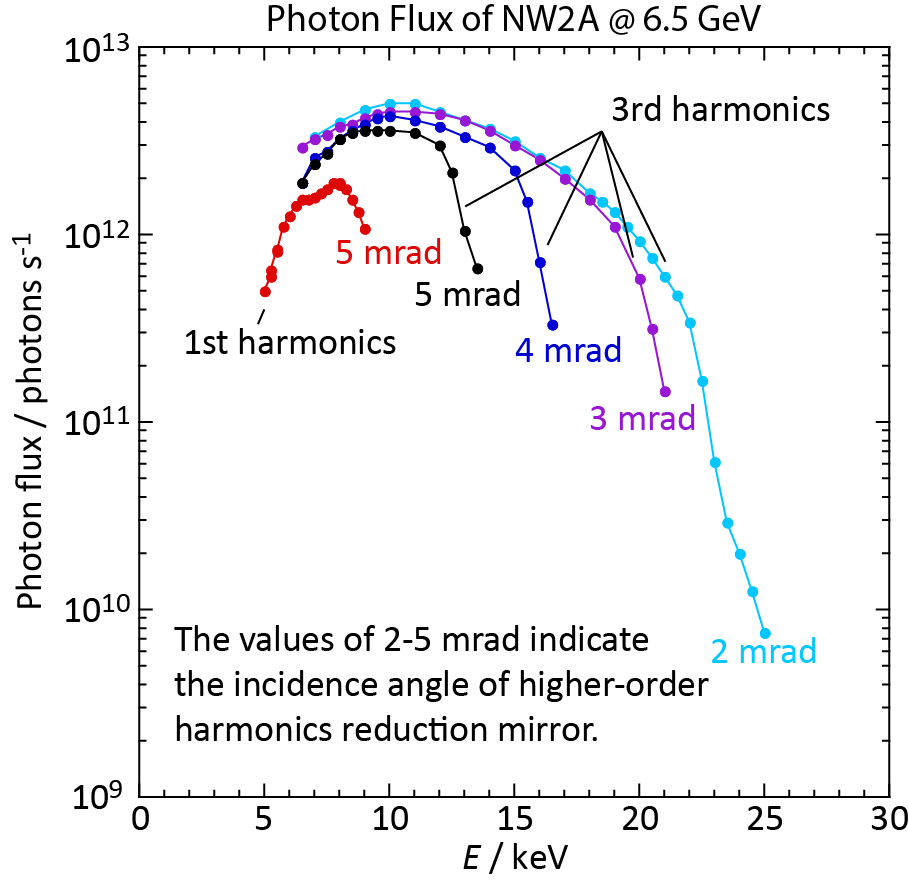
PF-AR Insertion device ID#2
- Ring energy: 6.5 GeV
- Max. current: 60 mA (Not Top-up operation)
- Critical energy: 26.3 keV
- Horizontal acceptance: 1 mrad(H) / 0.2mrad(V)
- Tapered mode available
Beamline optics
Monochromator
- Si(111) Liquid N2-cooled double crystal
- Energy range: 5 – 24 keV
- Numerical-drive (Kohzu system)
Focusing system
- Bent cylinder mirror (Rh coated)
- Bent flat mirror (Rh coated)
- Standard beam size: 0.6H x 0.2V mm
Focused and Monochromatized mode
- Energy range: 5 – 24 keV
- Energy resolution: ΔE/E~2×10-4
- Photon flux at sample position: 6×10¹² phs/s (12 keV)
White X-ray mode
Experimantal station
Experimental hutch
- Hatch size: 3.0L x 3.8W x 3.0H m
- Door size: 2.6W x 2.4H m
Standard setup
- XAFS setup: Ionization chumber
- DXAFS setup: Polychromator, PDA detector
- 2D Imaging XAFS setup: XAFS stage, ORCA Flash 4.0
- X-CT: Xradia 800 Synchrotron (ZEISS)
Beamline manager
- 丹羽 尉博 NIWA Yasuhiro (KEK-PF)
yasuhiro.niwa[@]kek.jp